Examples of X-ray optical component development
For those looking for a manufacturing partner in X-ray optics research
For many years, NTT-AT has assisted synchrotron researchers with the development of X-ray optical components. Here, we would like to introduce some of our X-ray optical components that have not been published as products.
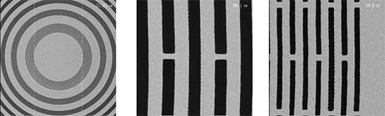
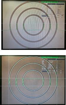
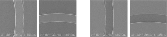
Case 1. Apotization X-ray Fresnel zone plate
Fresnel zone plate with improved resolution through continuously varying absorber thickness.
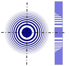
In X-ray microscopes that use synchrotron radiation as a light source, Fresnel zone plates (FZPs) are generally used as imaging elements.
NTT-AT has developed an "apodization FZP" in which the thickness of the X-ray absorber is continuously changed from the inside to the outside.
By using this FZP, it becomes possible to realize an X-ray microscope that achieves both high light-gathering efficiency and high resolution.
Furthermore, research results using this device were published in 2017 by Dr. Takeuchi's group at the Japan Synchrotron Radiation Research Institute (JASRI).
A Takeuchi et al, J. Phys.: Conf. Ser. 849 012055 (2017)
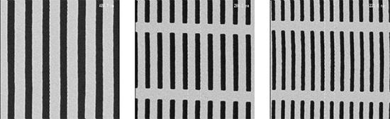
Case 2. Sector capacitor zone plate
Contributes to uniform illumination of the object being observed in X-ray imaging microscopes.
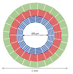
The sector condenser zone plate is an optical element developed for the illumination of X-ray imaging microscopes using synchrotron radiation.
This optical element has a structure in which equally spaced diffraction gratings are arranged radially, and by combining it with a center beam stop and an OSA (aperture for selecting the diffraction order of the zone plate) and rotating it, uniform illumination of the observation target can be obtained. can be realized.
NTT-AT 's sector condenser zone plate, in which three types of radially arranged transmission spacing diffraction grating patterns are arranged on a SiC membrane, was used for X-imaging microscopy experiments using SPring-8 BL37XU. Contributed to the realization of stable lighting.
In 2009, Dr. Takeuchi's group at the Japan Synchrotron Radiation Research Center published research results using this device.
A. Takeuchi et al, J. Phys.: Conf. Ser. 186 012020 (2009)
Case 3. X-ray Zernike phase ring
A transmission element for realizing an X-ray phase-contrast microscope.
The technique of converting the phase difference of transparent materials into light and dark contrast for detection is called phase difference observation and has various applications. A ring element that provides a phase difference of λ/4 is used for this phase difference observation.
In X-ray phase contrast observation, an X-ray Zernike phase ring is an element that imparts a phase difference of λ/4 to the X-rays.
NTT-AT 's Ta-based X-ray phase rings are mounted on synchrotron radiation-based X-ray imaging microscopes and are used for detecting microscopic phase-contrast images.
Furthermore, research results using this device were published in 2009 by Dr. Takeuchi's group at the Japan Synchrotron Radiation Research Institute (JASRI).
A. Takeuchi et al, J. Phys.: Conf. Ser. 186 012020 (2009)
inquiry
In addition to the products introduced here, NTT-AT provides a wide range of X-ray optical components, supporting our customers' research and development in fields such as synchrotron radiation science, laser applications, and industrial equipment. Please feel free to contact us for quotes and other inquiries.